
Figure 3.
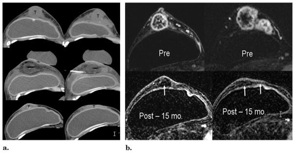
(a) CT guidance before and after cryotherapy. Top images, unenhanced CT images at the level of the nipple and superior breast show extensive tumor involvement (T). The skin and tumor appear inseparable but were NOT fixed at physical examination or involved at US or MR imaging (Fig 5b). Middle images, CT images at the same levels immediately after cryotherapy and probe removal, leaving air in tracts. The tumors are covered by hypodense ice, except for the image on right, where the ice had already melted after cryoprobes had been retracted from an earlier medial freeze cycle. The saline injection needle remains between the tumor and the implant and skin had also been infiltrated. Bottom images, CT images obtained at the same anatomic levels 15 months after cryotherapy show marked resolution of prior bulky tumors. Random biopsies performed six weeks after ablation showed no residual tumor (T), but cancer recurred in two sites beyond the ablation zone at seven months. Repeat cryotherapy was done for a 6-mm new nodule in the far upper central breast and a 17-mm lower axillary node. (b) MR evaluation before (Pre) and 15 months after (Post) cryotherapy. Top images, before cryotherapy, T1-weighted fat-suppressed gadolinium-enhanced axial MR images show brisk enhancement throughout the tumor nodularity of the retroareolar (left) and superior (right) breast. Note also the lack of skin enhancement. Bottom images, T1-weighted fat-suppressed gadolinium-enhanced MR images obtained at the same levels 15 months after cryotherapy show a marked reduction of prior bulky tumors and no residual enhancement of any margin (arrows).