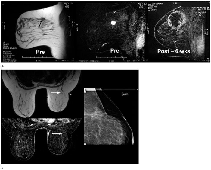
Figure 4.
(a) MR images obtained before (Pre) and six weeks after (Post) cryotherapy of multifocal left upper outer quadrant cancer. Left image, T1-weighted axial image shows the spiculated, 1.6-cm dominant mass in the left outer quadrant (arrow). Another biopsy-confirmed cancer in the same quadrant was seen in different image planes. Middle image, fat-subtracted T1-weighted image obtained after the administration of gadolinium shows brisk tumor enhancement. Right image, T1-weighted, fat-subtracted, gadolinium-enhanced MR image obtained six weeks after cryotherapy shows a rim of enhancement corresponding to a healing cryotherapy rim, which also covered the other cancer. The microlumpectomy sampling (ie, Mammotome removal) of the tumor region before initiation of the freeze made our first BC procedure more complex. US visualization of the tumor site became distorted by blood during the vacuum-assisted biopsies, which are no longer used. Despite poor visualization of any remaining tumor margin, thorough ablation margins were still achieved by placing the cryoprobes along the outer margins of these microlumpectomy sites. Biopsy of three slightly nodular areas of rim enhancement showed no residual cancer or DCIS. (b) Images obtained at five-year follow-up. Top left image, T1-weighted axial MR image shows minimal scarring and/or distortion at the cryosite (arrow). Slightly smaller left breast was initially present and lateral scar indentation (arrowhead) is a scar from abscess débridement performed one year before this study (ie, four years after cryotherapy), which also confirmed no residual tumor throughout the original cryotherapy site. Bottom left image, T1-weighted fat-subtracted, gadolinium-enhanced MR image shows no enhancement of the previous ablation and subsequent resection sites (arrow). Right image, compression mammogram of the left upper outer quadrant shows no mass effect and minimal scarring, likely related to resection one year ago rather than original cryotherapy five years ago.

