Figure 1.
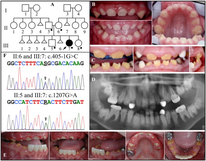
Family 1. (A) Pedigree. Dots mark the five persons who donated samples for DNA sequencing. Triangles represent stillborns. (B) Oral photographs of the proband (III:7) at age 2.5 yrs. (C) Oral photographs of the proband at age 8.5 yrs. The anterior incisors are present and exhibit severe enamel hypoplasia. The white cuspids are dental restorations. The attached gingiva is enlarged, but the impression is enhanced by the small sizes of the clinical crowns. (D) Panorex of the proband at age 11.5 yrs. Enamel is missing or does not contrast with dentin throughout. Most tooth roots are short, and eruption is less than expected. Pulp stones are observed in many teeth, particularly in the first molars. (E) Oral photographs of the proband at age 13 yrs. The gingival hyperplasia is minimal and could readily be missed in an oral examination. (F) DNA sequencing chromatograms of Family 1. Top: Sequence from the border of Exon 1 and Intron 1, revealing heterozygosity for a splice junction mutation (g.502011G>C; c.405-1G>C) that occurred in the mother and proband. Bottom: Exon 8 sequence revealing heterozygosity for a missense mutation (g.65094G>A; c.1207G>A; p.D403N) that occurred in the father and proband. The unaffected brother (III:5) and sister (III:8) had neither of these mutations (data not shown). The mutation designations are with respect to the FAM20A genomic reference sequence NG_029809.1 and cDNA reference sequence NM_017565.3 (for mRNA transcript variant 1). Key: arrowhead = mutation point; R = A or G; S = G or C.

