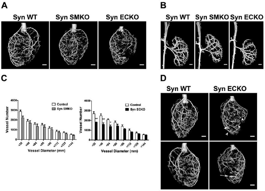
Figure 3. Developmental arteriogenesis defects are present in SynECKO but not in SynSMKO mice.
Representative reconstructed mCT images of whole hearts (A) and kidneys (B) arterial vasculature (16µm resolution; n=4) from age- and gender-matched SynWT, SynSMKO and SynECKO mice. Note marked reduction in branching in SynECKO mice compared with SynSMKO or SynWT control mice. (C) Quantitative analysis of mCT images of whole hearts (grey bars, SynSMKO and black bars SynECKO). Note a marked decrease in total number of <64µm diameter arterial vessels in SynECKO mice relative to control littermates.): F = 3.962, p = 0.003; post-hoc Tukey's HSD test: * p<0.01; ** p<0.001. (D) Reconstructed mCT images of the heart vasculature from SynECKO mice. Note the presence of aneurism-like structures (arrow) and increased diameters in the distal part of some arteries compared with their proximal partly (arrowheads). Scale bar 1mm.

