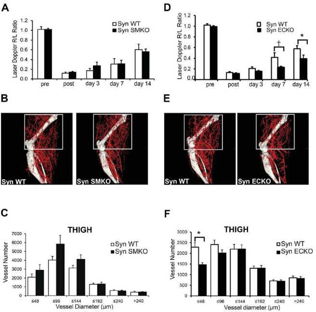
Figure 5. Impaired blood flow recovery in SynECKO mice following ligation of femoral artery.
(A, D) Laser Doppler analysis of blood flow perfusion: SynSMKO (A) and SynECKO (D) mice were subjected to common femoral artery ligation. The graph shows blood flow in the ischemic foot (right) expressed as a ratio to flow in the normal foot (left) (R/L) at various time points after femoral artery ligation. Note the absence of flow recovery in SynECKO mice at 7 and 14 days. Mean±SD, *p<0.05, †p=0.07, n=10 per group. (B, E) Representative mCT images of reconstructed limb arterial vasculature from SynSMKO (B) and SynECKO (E) compared with control littermates 14 days after surgery (C, F) Quantitative mCT analysis in SynSMKO (C) and SynECKO (F) compared with control littermates. Note a significant decrease in the amount of smaller size arteries in SynECKO mice compared with control group (C) *p<0.05, n=5 mice/group.

