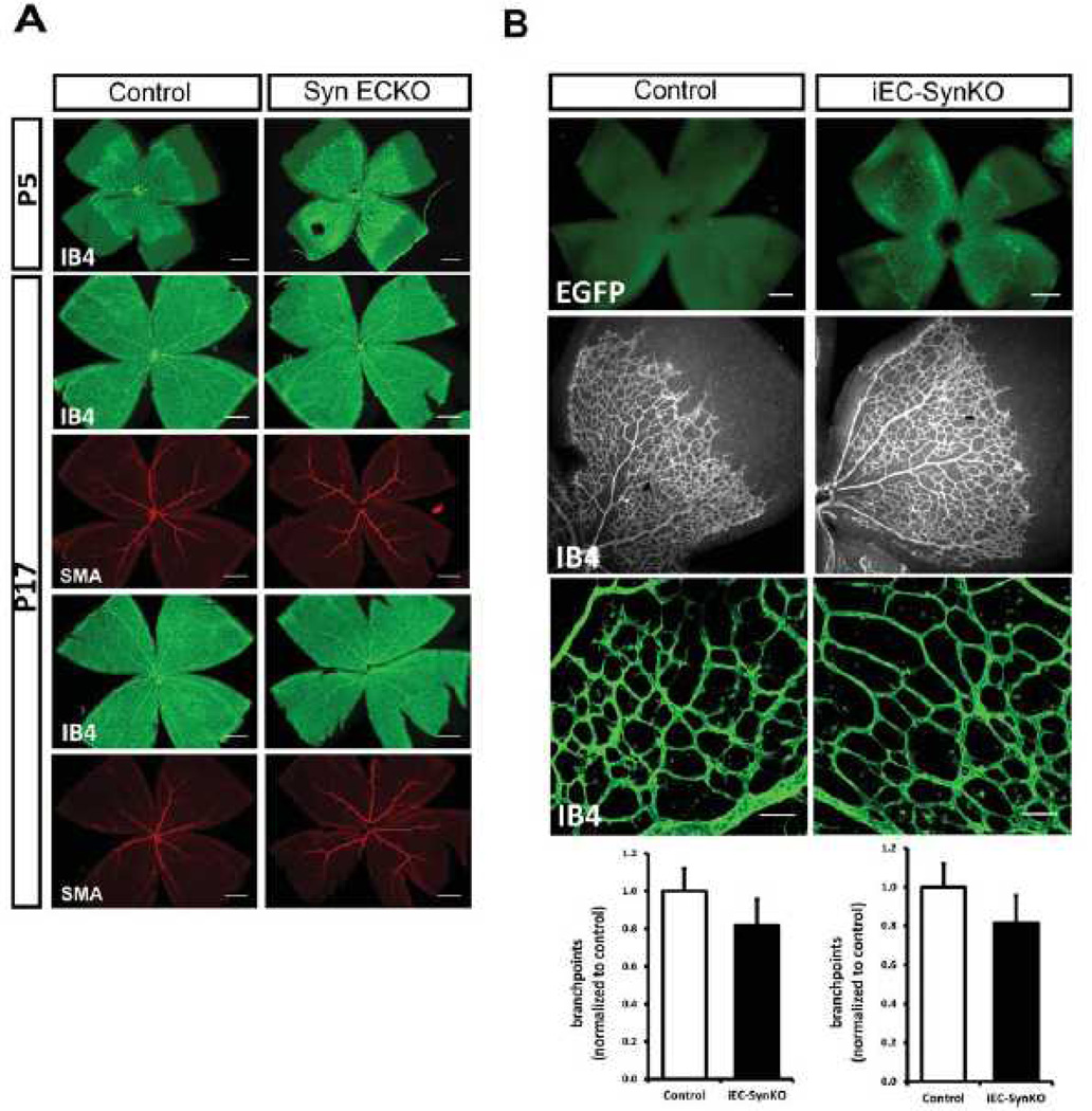
Figure 7. Retinal angiogenesis is not affected in SynECKO and iEC-SynKO mouse lines.
(A) Whole mounts of P5 and P17 retinas from SynECKO mice labelled with isolectin B4 (IB4) and stained with smooth muscle α-actin (SMA) antibody. Representative images for P5 (top) and P17 (two, bottom) are shown. There were no visual differences in vessel area and SMC coverage between SynECKO mutants and controls. Scale bar 0.5mm. n=4 mice/group. (B) Quantitative analysis of P5 whole mount retinas from iEC-SynKO mice generated with Pdgfb-iCre. Expression of EGFP in mutant pups demonstrates the efficiency of Cre induction after tamoxifen administration (top, scale bar 200 µm). Retinal vasculature and quantitative analysis of vascular area and vessel branchpoints are shown (bottom, scale bar 50 µm, N=3 mice/group.

