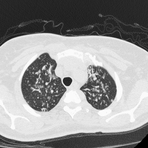
Figure 3.

High-resolution computed tomography image with 0.625 mm craniocaudal thickness and high spatial frequency reconstruction algorithm reveals subpleural location of some nodules. Pulmonary vascular margins are smooth, excluding perivascular nodules. The presence of subpleural nodules and the absence of beaded vascular margins argue for random nodular distribution due to hematogenous dissemination.
