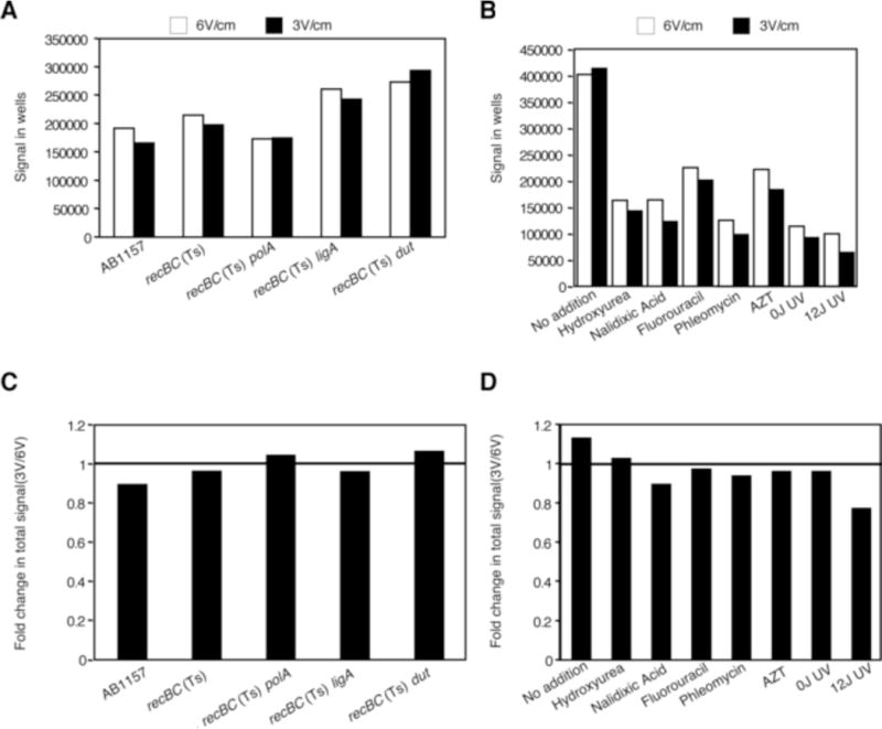
Figure 2.

Effect of electrophoretic conditions on the amount of signal staying within the wells (A and B); and total signal (well+lane) (C and D). Values are derived from the gels in Fig 1.
(A) Differences in the absolute amount of signal (arbitrary units) within wells in strains undergoing spontaneous fragmentation.
(B) Differences in the absolute amount of signal (arbitrary units) within wells in recBC(Ts) cells exposed to a variety of DNA damaging agents.
(C) Decrease in the total signal in strains undergoing spontaneous fragmentation, presented as ratios of the total signal in the LLFS conditions divided by the total signal from the same plug in the SHFS conditions.
(D) Decrease in the total signal in strains exposed to various clastogens (calculated as in “C”).
