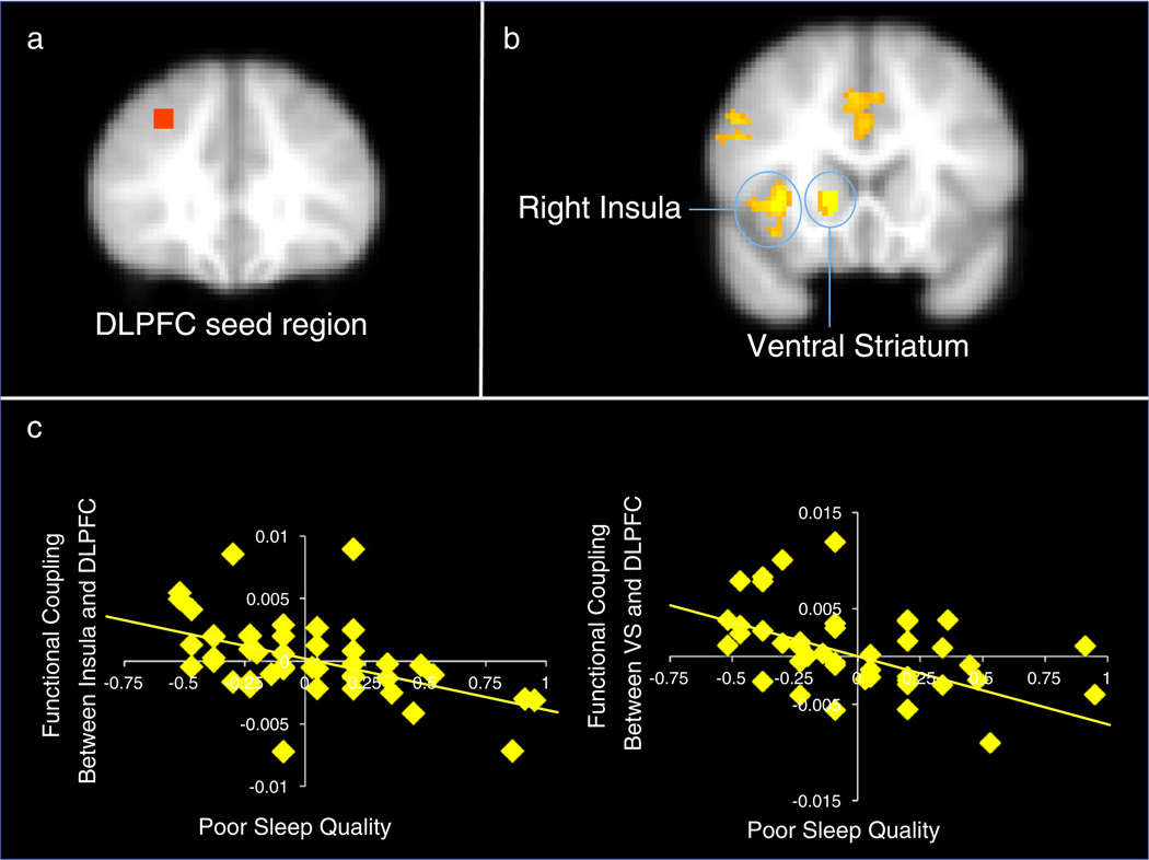
Fig. 5.
(a) the seed region was defined as a 6 mm sphere centered in the DLPFC from the cluster that correlated with sleep quality during the GNG. (b) Psychophysiological interaction analyses reveal less functional coupling between the DLPFC and neural regions for adolescents with poorer sleep quality. (c) Percent signal change in the insula and ventral striatum that showed decreased functional coupling with the DLPFC that correlated negatively with sleep quality. Note, greater values on the x-axis represent poorer sleep quality. Note. Right=left. Scatterplots are provided for descriptive purposes. All reported statistics are obtained from independent tests in FSL regressing sleep on brain activation.

