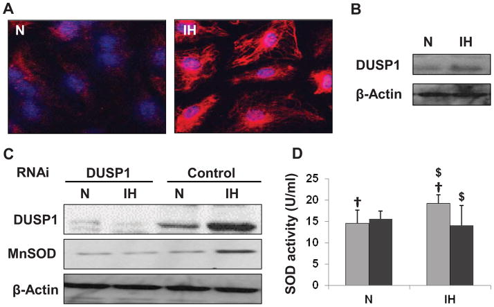
Figure 1.
Effect of IH treatment in human coronary artery endothelial cells (HCAEC) and granulocytes. (A) Confocal images showing increased expression and nuclear (blue) localization of DUSP1 (red) in intermittent hypoxia treated HCEAC. (B) Representative Western blot showing increased DUSP1 expression in cultured granulocytes after IH treatment. (C) Representative Western blots showing increased expression of DUSP1 and MnSOD in control-RNAi transfected cells after IH treatment. The cells transfected with DUSP1 RNAi failed to show any increase in DUSP1 or MnSOD after IH treatment. (D) Graphical representation of SOD activity in HCAEC cells from Control RNAi (light grey bars) and DUSP1 RNAi (dark grey bars) treated cells after IH treatment. IH increased SOD activity in control, however cells transfected with DUSP1 RNAi did not show any increase in SOD activity after IH exposure. Data presented as mean ± SD of three independent experiments. Statistical significance was determined by unpaired t-tests. †is P<0.05 N vs. IH treatment in control-RNAi transfected cells. $ is P<0.005 control vs. DUSP1 RNAi transfected cells in IH condition. N: Normoxia; IH: Intermittent hypoxia.

