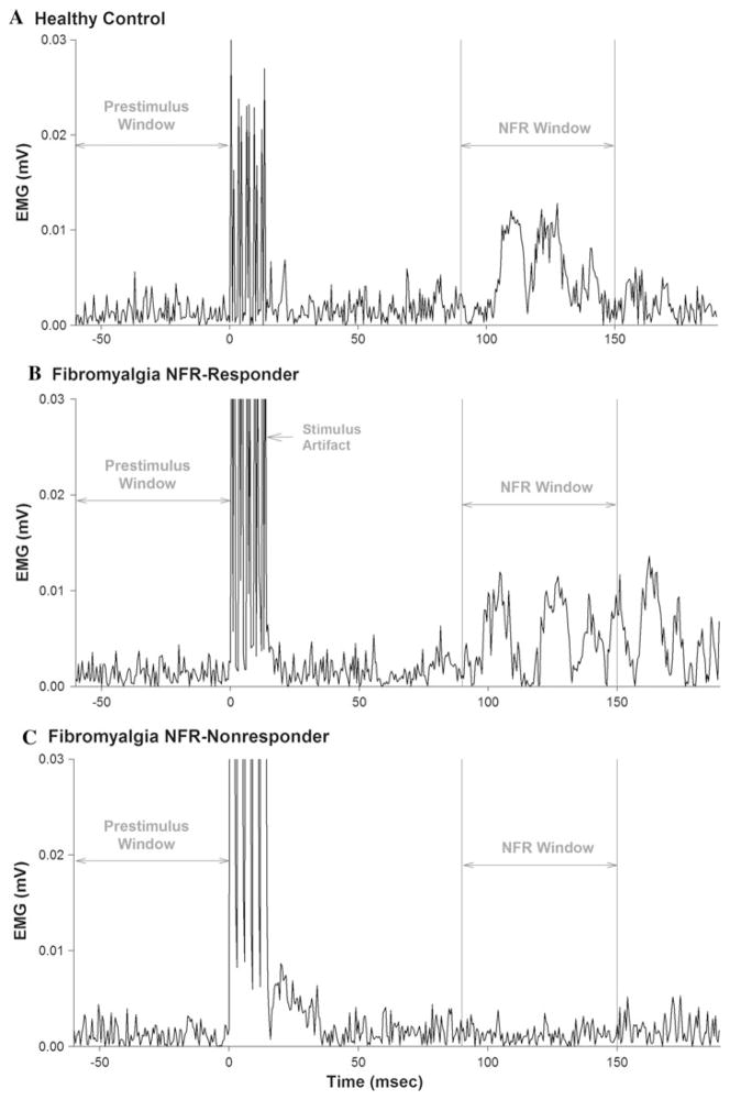
Fig. 2.
Nociceptive flexion reflexes (NFR) in the biceps femoris muscle are shown for representative women from the healthy control group (a), the NFR responder group (b), and the NFR nonresponder group (c). A positive NFR response was defined as an increase in the rectified electromyographic (EMG) signal within a time window corresponding to the NFR latency (90–150 ms after delivery of a train of electrical stimuli to the sural nerve) that exceeded the mean pre-stimulus EMG by ≥1 SD. Note that no NFR response was detected for the individual shown in (c) at the maximum stimulus intensity of 40 mA (relative stimulus intensity = 19 × perceptual threshold)

