Figure 2.
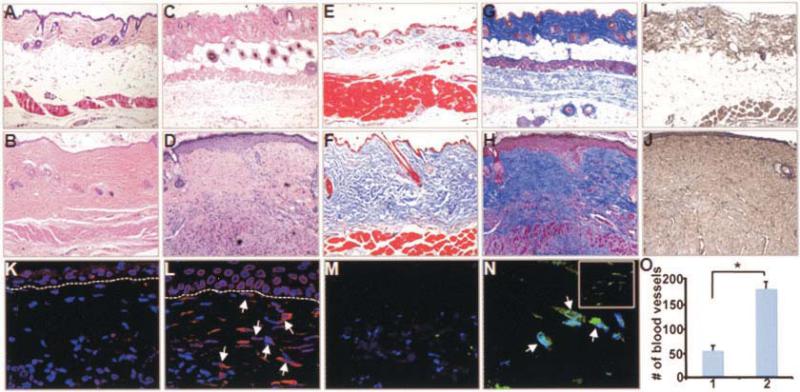
Extensive dermal fibrosis in adult Col1a2-CTGF–transgenic mice. A–N, Skin biopsy samples from WT littermate controls (A, C, E, G, I, K, and M) compared with those from adult Col1a2-CTGF–transgenic mice (B, D, F, H, J, L, and N) at 4 weeks of age (A, B, E, and F) and 8 weeks of age (C, D, G–O). Histologic results are representative of skin biopsy samples from 10 mice per group. Compared with WT mice, Col1a2-CTGF–transgenic mice showed pronounced and progressive dermal fibrosis with focal thickening of the epidermis, by hematoxylin and eosin staining (A–D) and Masson's trichrome staining (E–H). Immunochemistry analysis revealed increased accumulation of type I collagen in sections from Col1a2-CTGF–transgenic mice compared with those from WT mice (I and J). Immunofluorescence with CTGF antibody revealed increased CTGF-expressing fibroblasts in Col1a2-CTGF–transgenic mouse skin sections (arrows) compared with WT mouse skin sections (K and L). Increased numbers of myofibroblasts were observed in the papillary and reticular dermis (N and inset in N, respectively) of Col1a2-CTGF–transgenic mouse skin (arrows) compared with WT mouse skin (M). O, Angiogenesis in WT mice (column 1) and Col1a2-CTGF–transgenic mice (column 2), quantified based on numbers of blood vessels. Values are the mean and SD. * = P < 0.001. See Figure 1 for definitions.

