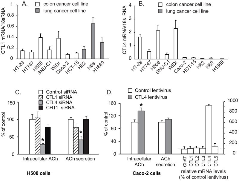
Fig. 5.
CTL4 modulates ACh synthesis in colon cancer cells. A. Expression of CTL1 in colon and lung cancer cell lines. B. Expression of CTL4 in colon and lung cancer cell lines. C.Effect of knockdown of CTLs 1, 4 and CHT1 on ACh synthesis of H508 cells. Specific siRNAs decreased levels of CTL1 and CTL4 mRNA respectively by 50–60% compared to control siRNA-treated cells (data not shown, but similar knockdown and specificity as shown in figure 3). Data are mean ± SE of 7 replicates in two separate experiments and show percent of control intracellular ACh (0.07 pmol by 100,000 H508 cells treated with control siRNA) or percent of control media ACh concentration (0.14 pmol per 100,000 H508 cells treated with control siRNA in 48 h). *p < 0.01versus control siRNA. 50 μM neostigmine added to all cultures to prevent ACh degradation. D. CTL4 transduction increased ACh synthesis by Caco-2 cells. Transduction of CTL4 significantly elevated intracellular ACh of Caco-2 cells by 35%, but did not significantly impact ACh secretion. Data are mean ± SE of 10 replicates in two separate experiments and presented as percentage of Caco-2 cells ACh secretion by cells transduced with control lentivirus or ACh secretion (1.76 pmol by 100,000 Caco-2 cells within 48 h) or percent of intracellular ACh in cell cells transduced with control lentivirus (3.91 pmol in 100,000 Caco-2 cells). 50 μM neostigmine added to all cultures to prevent ACh degradation. Right hand bars show that CTL4 transduction increased CTL4 mRNA levels 9.1-fold compared to cells transduced with control lentivirus, but did not significantly affect levels of the other CTL or ChAT mRNA. *p < 0.01 versus control lentivirus.

