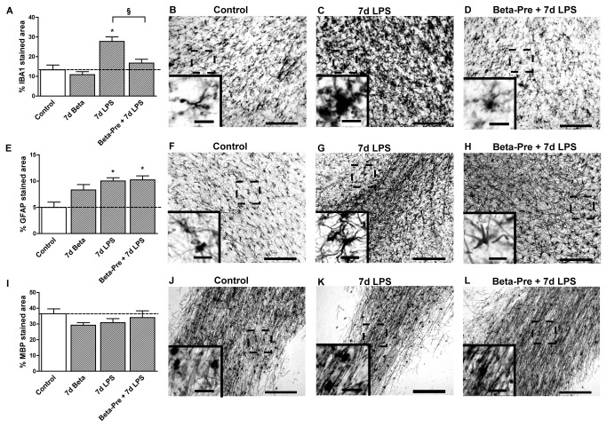
Figure 3. Effect of intra-amniotic LPS and antenatal glucocorticoid exposure 7 days before delivery on the subcortical white matter.
A: The area fraction (%) of IBA1 immuno-reactivity in the subcortical white matter increased significantly after intra-amniotic LPS exposure 7 days before delivery. B-D: Representative images of the IBA1 staining in the SCWM in controls (B), 7d LPS (C) and Beta-Pre + 7d LPS (D) exposed animals. E: LPS exposure 7 days before delivery significantly increased the area fraction (%) of GFAP immuno-reactivity in the SCWM. F-H: Representative images of the GFAP staining in the SCWM in controls (F), 7d LPS (G) and Beta-Pre + 7d LPS (H) exposed animals. I: The area fraction (%) of MBP immuno-reactivity did not differ in animals exposed to intra-amniotic LPS and/or betamethasone exposure 7 days before delivery. J-L: Representative images of the MBP staining in the SCWM in controls (J), 7d LPS (K) and Beta-Pre + 7d LPS (L) exposed animals. Scale bar = 200 µm; scale bar insert = 25 µm. *p<0.05 versus controls and § p<0.05 between experimental groups using a one-way ANOVA with Tukey’s post hoc test.

