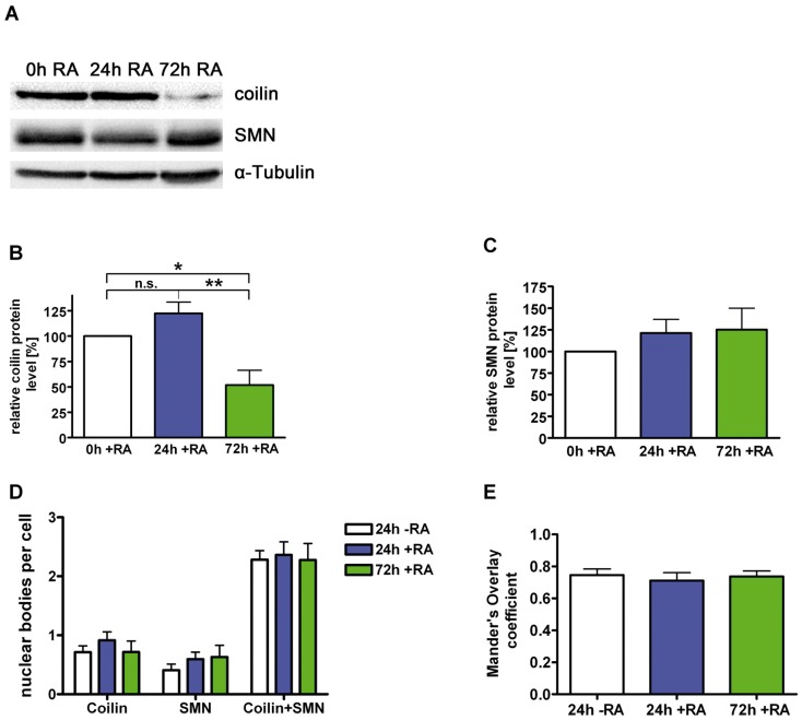
Figure 3. Regulation of the SMN-interacting protein coilin.
Neuroblastoma SK-N-BE(2) cells were differentiated for different periods with retinoic acid (5 µM), lysed by sonification in modified RIPA-buffer [7] and analyzed by Western blot for coilin, SMN and α-Tubulin (A). (B) Endogenous coilin protein levels were decreased significantly to 52% after 72h of differentiation, (C) while SMN protein levels were increased non-significantly after the same time of differentiation. (D) Cells were treated with RA and stained with anti-SMN and anti-coilin antibodies. Coilin-positive (SMN-negative Cajal bodies), SMN-positive (nuclear gems) as well as coilin- and SMN-positive dots (SMN-positive CBs) in the nucleus of differentiated NB cells were counted and compared with a non-differentiated control. No significant changes of absolute numbers of these nuclear bodies were detected. (E) Intensity correlation analyses of coilin and SMN in the nucleus of differentiated cells were performed and compared to a non-differentiated control. No change in colocalization of coilin and SMN was detected (B, C: n = 7, means ± SEM; *, p<0.05; **, p<0.01; unpaired, two tailed t-test; D: control, n = 18; 24h RA, n = 13; 72h RA, n = 20; E: control, n = 20; 24h RA, n = 15; 72h RA, n = 22).

