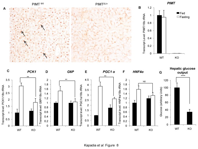
Figure 8. Ablation of PIMT expression in liver reduces gluconeogenesis.
(A) De-paraffinized 5 μm thin sections were immunohistochemically stained using PIMT antibody in WT and KO mice of liver tissue. (B) Total RNA was isolated from liver tissue and qPCR analysis of PIMT in PIMTfl/fl (WT) and PIMTΔLiv (KO) livers was performed. (C-F) WT and KO mice were fasted for 72h. RNA was extracted and qPCR was performed for PCK1 (C), G6P (D), PGC1α (E) and HNF4α (F). (G) Primary hepatocytes were isolated from WT and KO mice. Post 24h of isolation, cells were culture in glucose production medium for 6 h. Amount of glucose released was estimated and the values were normalized to the corresponding protein content. The values are expressed relative to the normalized WT control group. Data are representative of three independent experiments. Statistical analysis was performed using unpaired Student’s t-test **p<0.01, ****p<0.001, NS: Non-Significant.

