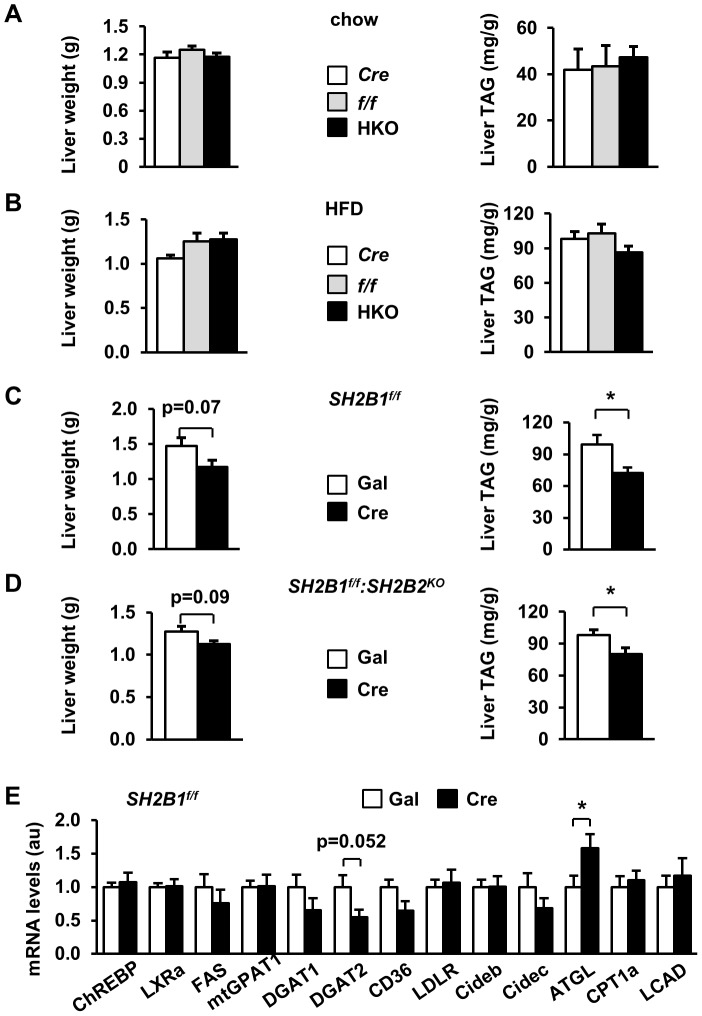
Figure 7. Hepatocyte-specific deletion of SH2B1 attenuates HFD-induced hepatic steatosis.
(A) Liver weight and liver TAG levels (normalized to liver weight) of overnight-fasted male mice (21 weeks of age). f/f: n = 10; Cre: n = 7; HKO: n = 10. (B) Male mice (7 weeks of age) were fed a HFD for 14 weeks. Liver weight and TAG levels of overnight-fasted mice. f/f: n = 10; Cre: n = 7; HKO: n = 10. (C) SH2B1f/f male mice (7 weeks of age) were fed a HFD for 5 weeks and infected with β-gal (n = 7) or Cre adenoviruses (n = 6) via tail vein injection. Liver weight and TAG levels were measured in fasted mice 29 days after adenoviral infection. (D) SH2B1f/f:SH2B2KO male mice (7 weeks of age) were fed a HFD for 5 weeks and infected with β-gal (n = 6) or Cre adenoviruses (n = 6). Liver weight and TAG levels were measured in fasted mice 29 days after adenoviral infection. (E) Total liver mRNA was prepared from the mice described in (C) and used to measure mRNA levels of the indicated genes by qPCR and normalized to β-actin expression. The values were further normalized to the β-gal group. Data are presented as means ± SEM. P<0.05.

