
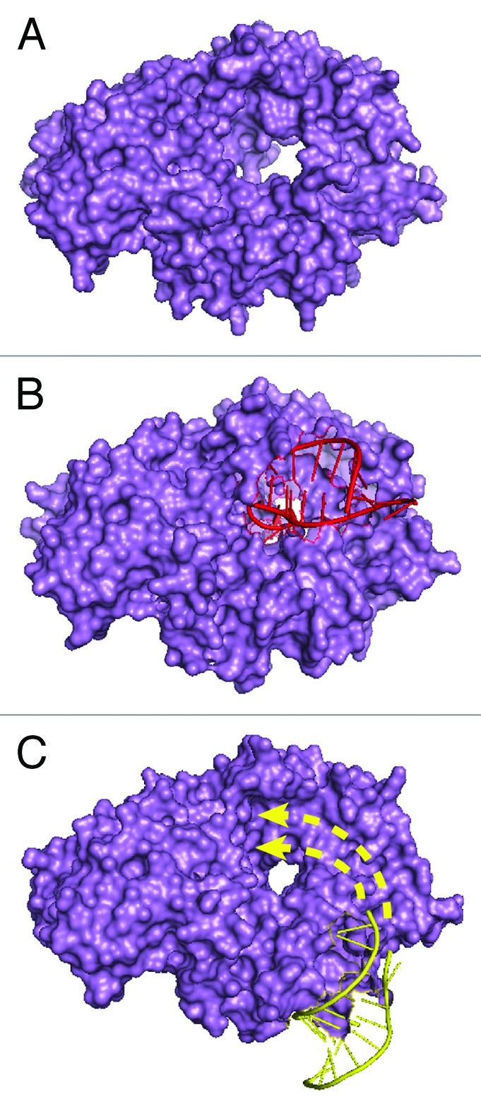
Figure 3. Structures of Ro60. (A) Molecular surface representation of X. laevis Ro60. The hole is 10–15 Å in diameter and binds single-stranded RNA.15 (B) X. laevis Ro60 bound to a misfolded pre-5S rRNA fragment consisting of a short duplex and a single-stranded 3′ extension. The duplex binds on the Ro outer surface, while the single-stranded end inserts through the cavity.27 (C) X. laevis Ro60 bound to a Y RNA fragment.15 The sequence used for crystallization is shown in bold in Figure 1B. Studies of Y RNA binding to mutant Ro60 proteins15,27 suggest that the remainder of the RNA interacts with portions of Ro60 that overlap the misfolded RNA binding site (arrows).