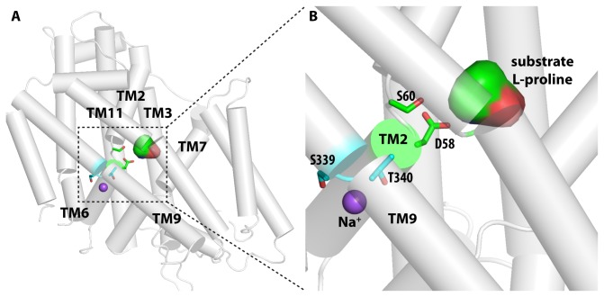
Figure 4. Homology model of HpPutP.
(A) Overview of the homology model of HpPutP. The substrate binding site is enclosed by TMs 2, 3, 7, and 11, while the bound Na+ ion is coordinated by residues from TMs 2, 6, and 9. (B) The zoom-in view of the predicted Na+ and L-proline binding sites. The substrate and Na+ binding residues that have been mutated (see text) are shown in stick representation.

