
Figure 1.
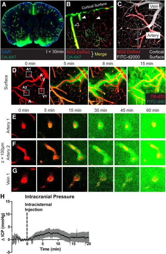
CSF enters the brain along para-arterial pathways. A, Fluorescent tracer (OA-647; MW, 45 kDa) injected intracisternally into the subarachnoid CSF enters the brain parenchyma along paravascular pathways. B, Ex vivo confocal imaging in NG2-DsRed mice showed that 15 min after injection, CSF tracer enters the brain along DsRed-positive penetrating arteries (hollow arrowheads), but not along DsRed-negative ascending veins (filled arrowheads). C, In vivo two-photon imaging in NG2-DsRed mice after fluorescence angiography (intravenous FITC-conjugated dextran-2000; MW, 2000 kDa) shows that cortical arteries and veins can be readily distinguished in these mice. D–G, Time-lapse in vivo two-photon imaging of florescent CSF tracer (FITC-d40; MW, 40 kDa) influx into the cortex after intracisternal injection. Cerebral vasculature is imaged by intra-arterial injection of Texas Red-conjugated dextran-70 (TR-d70; MW, 70 kDa). D, At the cortical surface, CSF tracer moves via para-arterial spaces. E, F, 100 μm below the cortical surface, CSF tracer enters the cortex along penetrating arteries, then exchanges with the surrounding interstitium. G, After para-arterial influx, CSF tracer is also evident along ascending veins. H, Measurement of ICP during intracisternal CSF tracer infusion (1 μl/min for 10 min) resulted in mild (∼2.5 mmHg) elevation of ICP that resolved rapidly upon cessation of infusion.