
Figure 2.
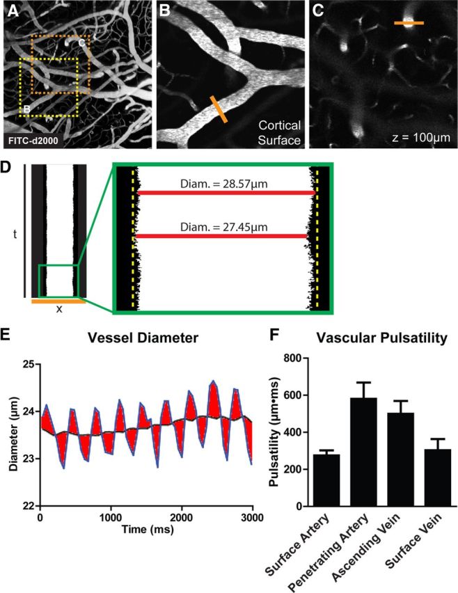
Measurement of vascular pulsatility by in vivo two-photon imaging. A, The cerebral vasculature was visualized by in vivo two-photon fluorescence angiography after intravenous injection of FITC-conjugated detran-2000 (FITC-d2000; MW, 2000 kDa). B, C, Cortical surface arteries and veins (B), and penetrating arteries and ascending veins (C) were selected and X–T line scans (orange lines) were generated orthogonal to the vessel axis. D, E, Vessel diameter was measured and plotted as a function of time. Vascular pulsatility was defined as the absolute value of the integral of the vascular diameter approximately a running average over a 3000 ms epoch. F, Vascular pulsatility was measured in cortical surface arteries (SA), penetrating arteries (PA), ascending veins (AV), and surface veins (SV). Pulsatility was greatest in penetrating arteries and veins compared with surface vessels.