
Figure 2.
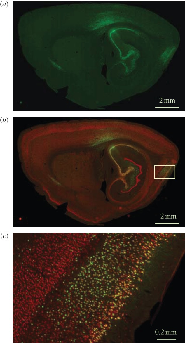
Sagittal sections showing viral transduction of hippocampus-projecting neurons in MEC. EYFP-carrying rAAV was injected in the dorsal hippocampus. (a) EYFP expression at low magnification (green), sagittal section; (b) section co-stained for NeuN (red), low magnification; (c) high magnification of the framed area in (b). The framed area shows the dorsal part of MEC. Note co-expression of EYFP and NeuN in layer II–III but not layer V–VI cells of the MEC. Modified with permission from [27].