
Figure 4.
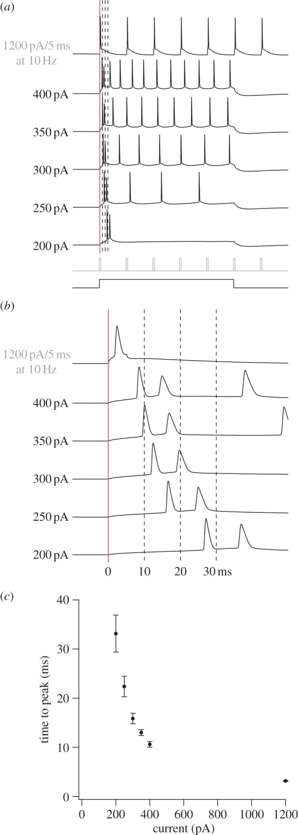
Firing latencies of stellate cells in MEC following intracellular current injection. (a) Series of whole-cell current steps in a P42 layer II stellate cell. Schematic of the 500 ms/5 ms current steps is shown below the data (500 ms for 200–400 pA; 5 ms at 10 Hz for 1200 pA). (b) Expanded view of 60 ms segment where vertical lines correspond to those also in (a). Red line indicates start of current step. (c) Graph of mean time to peak values (±s.e.m.) as a function of current, derived from 16 adult stellate cells (P31–P49).