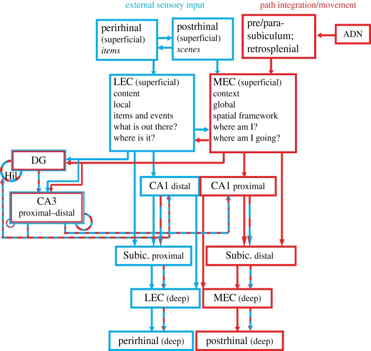
Figure 1.
Parallel processing streams into the hippocampus. This wiring diagram is a simplified version of the real anatomy, leaving out a number of projections. The structure of the diagram emphasizes the dual processing streams that pass through the LEC and MEC. Prior diagrams of these processing streams stressed their origins in the perirhinal-LEC and postrhinal-MEC connections [5–8]. Here, we add the critical connectivity between the MEC and limbic regions involved in movement, location and head direction processing (presubiculum, parasubiculum, retrosplenial cortex and anterior dorsal thalamus). (A different subregion of the parasubiculum projects to the LEC, not shown on this diagram [9].) The LEC and MEC connect to distinct regions of CA1 and subiculum, segregated along the transverse axis of the hippocampus (proximal–distal relative to the DG). CA1 and subiculum send return projections to the deep layers of the entorhinal cortex (EC), completing a processing loop. There is crosstalk along these pathways, both prior to their entry into the hippocampus and especially in the convergent projections to the DG and CA3. Although this article does not discuss DG and CA3 properties in detail, these areas are included in the diagram because they are major components of the classic ‘trisynaptic loop’ circuit of the hippocampus and it is important to place this circuit within the larger context of the MEC–LEC parallel streams. In this illustration, the DG and CA3 is represented as a ‘side loop’ of processing, in which the MEC and LEC streams are merged onto the same CA3 pyramidal cells and DG granule cells and the combined representations are then merged in CA1 with the separate input streams from the direct EC–CA1 projections. Specific mnemonic properties of the DG and CA3 regions are thought to be supported by the recurrent feedback loops represented by the dashed circles. In CA3, the recurrent connections are more prominent in the distal than the proximal regions [10]; moreover, the distal CA3 projects more strongly to proximal CA1 (which receives MEC input), and proximal CA3 projects more strongly to distal CA1 (which receives LEC input). The DG receives feedback from CA3 [11], and a disynaptic recurrent loop via mossy cells of the hilus is also present [12] For a more detailed explanation and references to the primary literature on these anatomical connections, the reader is referred to a number of review articles [5,13–17]. ADN, anterior dorsal nucleus of the thalamus; DG, dentate gyrus; Hil, hilus; Subic., subiculum. (Online version in colour.)

