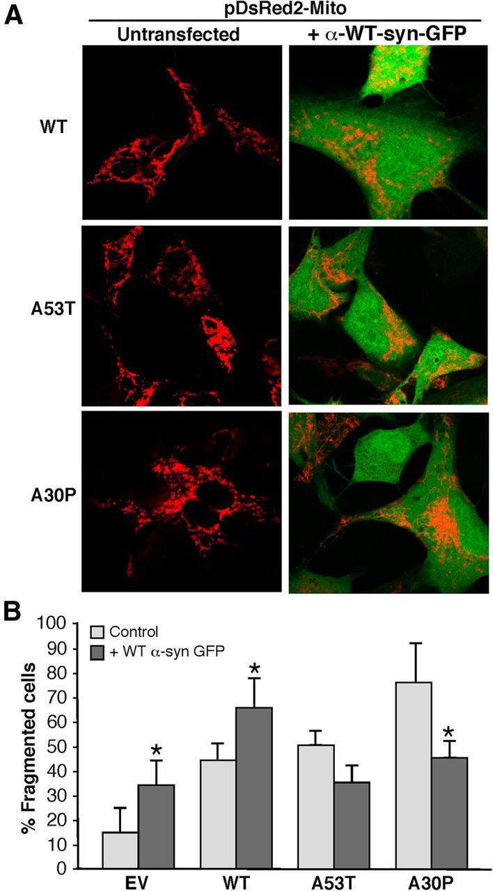
Figure 12.

Relationship of WT α-syn expression to mitochondrial fragmentation. A, M17 cells stably transfected with an EV plasmid or with the indicated plasmids expressing α-syn species were transiently cotransfected with plasmids expressing pDsRed2-Mito (red) and WT α-syn-GFP (green) and visualized by confocal microscopy. Cells were inspected visually and scored for whether they contained predominantly tubular or predominantly fragmented mitochondria (red). Note that, compared with Figure 7, the fragmented phenotype in the α-syn mutant cells is ameliorated by the expression of WT α-syn-GFP. B, Quantitation of percentage fragmented cells shown in A. *Significant difference versus corresponding untransfected cells (p < 0.05).
