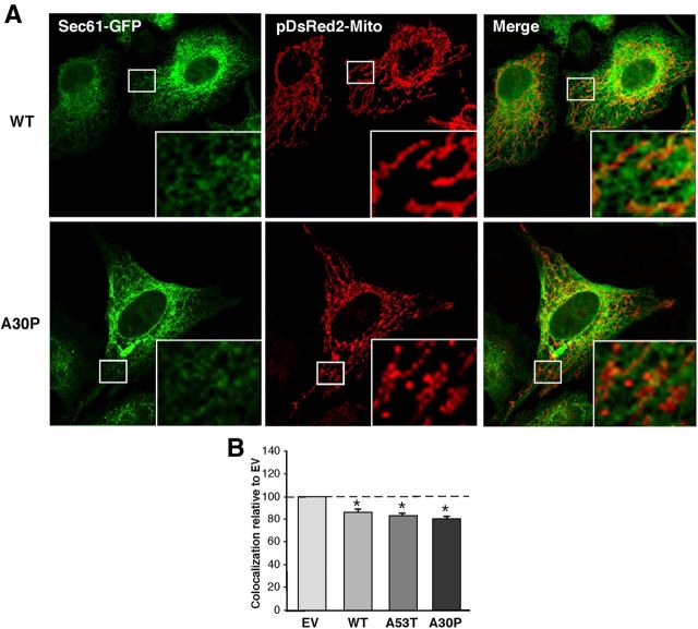
Figure 5.
Apposition of ER and mitochondria in HeLa cells. A, Examples of colocalization of ER labeled with GFP-Sec61-β (green), and mitochondria labeled with pDsRed2-mito (red), in HeLa cells transiently expressing WT- and A30P-α-syn. B, Quantitation of colocalization (as in A) by ImageJ analysis, normalized to the baseline level of apposition value in EV cells (baseline = 100; average of 4 experiments ±SD). Asterisk denotes significance versus EV.

