
Figure 3.
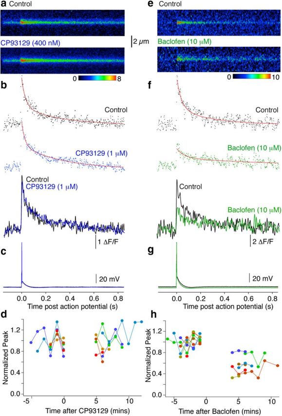
Differential inhibition of Ca2+ transients by 5-HT1BRs and GABABRs. a, Line scans (as in Fig. 2) from individual varicosities in the subiculum to resolve action potential-evoked (c) presynaptic Ca2+ transients (mean 3 responses in Control and 3 in CP93129; 1 μm). b, Signals were integrated for the sweeps over the terminal varicosities and amplitudes expressed as ΔF/F (Control, black; in CP93129, blue). Peak amplitudes were calculated from double exponential fits (b, red). Traces overlaid (below) show no effect of CP93129 application (time base identical, a–c). d, Peak amplitudes normalized to the mean Control amplitude before drug application were plotted against time for all cells. Each data point is the response to a single action potential in a single terminal varicosity. Colors represent results from individual terminals (n = 6). e–g, In another neuron baclofen (10 μm) imaged and stimulated under the same conditions as above significantly reduced Ca2+ transient amplitudes. h, Amplitudes of individual action potential-evoked Ca2+ transients demonstrate that in all seven neurons, although individual responses were reduced in amplitude in baclofen, failure of responses never occurred.