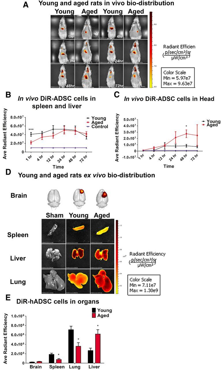
Figure 4.

Biodistribution of DiR-labeled hADSCs after TBI. In vivo near-infrared IVIS imaging revealed that the biodistribution and homing of hADSCs is age dependant. A and B show that hADSCs migrated robustly to the spleen and the liver area of young rats exposed to TBI relative to sham controls and to the spleen and liver area of aged rats exposed to TBI and transplanted with hADSCs (one-way ANOVA, F(3,9) = 6.7, p < 0.001; Bonferroni's test, ***p < 0.001). C shows that the migration signal is significantly higher in aged rats only within the first 48 h compared with young and sham control (one-way ANOVA, F(3,9) = 5.61, p < 0.05; Bonferroni's test, *p < 0.05). The migration signal of the labeled hADSCs to the brain of young rats exposed to TBI was low and reached a plateau at 12 h after transplantation compared with aged rats and sham controls. D shows ex vivo near-infrared IVIS imaging of the brain, spleen, lungs, and liver 11 d after TBI. E, Ex vivo fluorescent analysis revealed that spleen and lungs of young rats exposed to TBI expressed higher migration of hADSCs compared with the spleen and lungs of rats exposed to TBI or sham controls; two-way ANOVA, F(3,9) = 14.15, p < 0.001; Bonferroni's test, *p < 0.05). Radiant Efficiency, photons per second per square centimeter per steridian divided by microwatts per square centimeter .
