Figure 1.
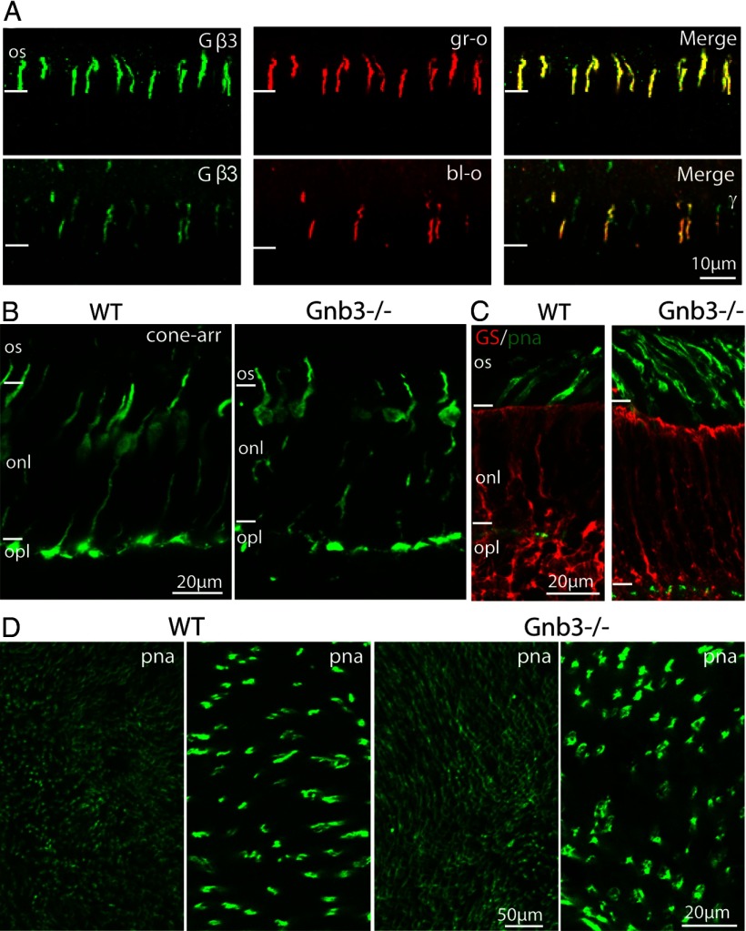
All cones express Gβ3 and Gnb3-null cones appear normal. A, Coimmunostaining for Gβ3 and green (gr-o, top) or blue (bl-o, bottom) opsin in a WT retina shows strong staining for Gβ3 in cone outer segments expressing either opsin. B, Immunostaining of WT and null (Gnb3−/−) retina with the cone-specific marker, cone arrestin (cone-arr), shows that Gnb3−/− cones have normal appearance and normal expression pattern of cone arrestin. C, Immunostaining of WT and Gnb3−/− retina for glutamine synthetase (red), shows similar staining pattern in the two genotypes, indicating absence of Muller cell proliferation in Gnb3−/−. Cone outer segments labeled with PNA (green) show normal morphology in the Gnb3−/−. D, Whole-mount retina staining for PNA shows normal cone density in the null retina (left two panels show WT at low and high magnification respectively; right two panels show KO). Three- to 8-week-old mice were used for these experiments. For this and subsequent figures: all immunostaining experiments were performed on at least 3 sets (KO and WT), and the same imaging parameters were used for a set. Os, Outer segments; onl, outer nuclear layer; and opl, outer plexiform layer.

