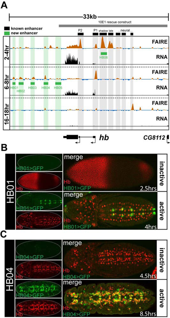
Figure 2. FAIRE signal accurately predicts enhancer activity.
(A) FAIRE and RNA signals at the hunchback (hb) locus in embryos. Black boxes designate the locations of known enhancers: (left to right) P2 promoter, P1 promoter, blastoderm shadow enhancer, late blastoderm enhancer, and recently identified neural enhancers (Gallo et al., 2011; Hirono et al., 2012; Margolis et al., 1995; Perry et al., 2011). Green boxes designate enhancers that were identified and cloned in this study. The grey box indicates the boundaries of the 10E1 transgenic hb rescue construct (Margolis et al., 1995). (B, C) Confocal images of embryos from two transgenic lines (HB01, HB04) stained with antibodies for Hb (red) and GFP (green) protein. The estimated age of each embryo is indicated. The timing of chromatin opening coincides with timing of reporter activity. See also Table S1 and Data File S1.

