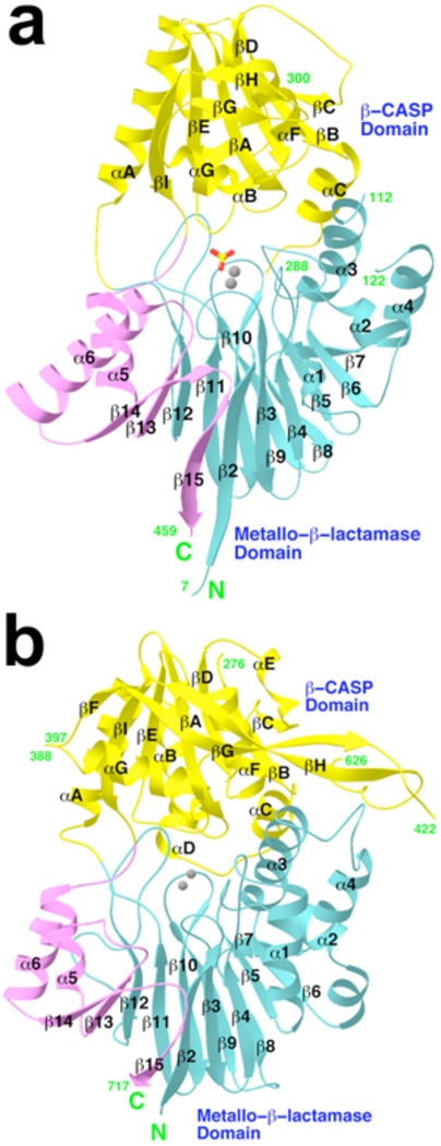
Figure 1. Structures of human CPSF-73 and yeast CPSF-100 (Ydh1p).
a, Schematic representation of the structure of human CPSF-73. The β-strands and α-helices are labeled, and the two zinc atoms in the active site are shown as gray spheres. The sulfate ion is shown as a stick model. b, Schematic representation of the structure of yeast CPSF-100. The zinc atoms in the CPSF-73 structure are shown for reference.

