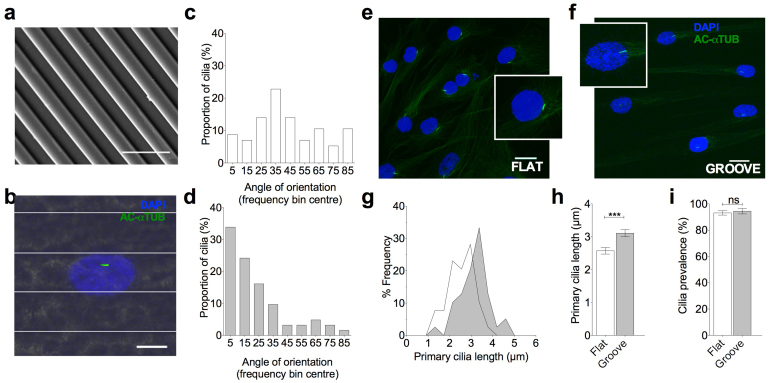
Figure 1. Stem cell primary cilia orientate parallel to substrate grooves and elongate compared with flat culture.
(a) SEM image of grooved substrate. Scale bar represents 10 μm. (b) Representative immunofluorescent confocal maximum projection image of MSCs cultured on grooved substrate. The primary cilia are labeled with anti-acetylated alpha tubulin (green) and nuclei counterstained with DAPI (blue). Scale bar represents 15 μm. Fluorescent image overlaid on reflectance image to enable marking of groove ridges. (c/d) Frequency distributions showing the angle of orientation of the primary cilia on flat and grooved substrates respectively. (e/f) Confocal 3D maximum projection images showing primary cilia in cells grown on flat and grooved surfaces respectively. Inserts showing primary cilia at 4 × higher magnification. Cells were labeled with anti-acetylated alpha tubulin (green) and nuclei counterstained with DAPI (blue). Scale bar represents 10 μm. (g) Frequency histogram for primary cilia length on flat and grooved surfaces (dark grey is the grooved surface).(h) Effect of topography on primary cilia length. Values represent mean with error bars indicating SEM. (i) Effect of topography on primary cilia prevalence with error bars indicating S.E.M.

