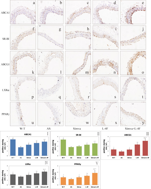
Figure 5.
Immunochemical staining of ABCA1, SR-BI, ABCG1, LXRα and PPARγ in abdominal aortic. a-y representative light microscopy images of abdominal aorta (X400). a-e ABCA1 staining; f-j SR-BI; k-o ABCG1; p-t LXRα; u-y PPARγ.I-V statistical results of positive staining (n = 6). 1P < 0.05, 2P < 0.001, vs. AS group; aP < 0.05, bP < 0.001, vs. Simva group;*P < 0.05, **P < 0.001, vs. L-4F group.

