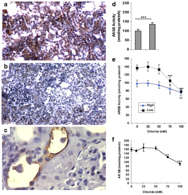
Fig. 1.
Reduced ARSB activity in vivo following higher salt exposure. a,b,c. Immunohistochemistry of representative renal tissue from SSL and SSH rats demonstrated increased intensity and extent of positive staining for ARSB in the epithelial cells of the proximal and distal tubules in the SSL renal tissue (a), compared to the SSH (b) renal tissue. Findings are consistent with reduced ARSB activity following exposure to high salt in the salt-sensitive rats (original magnification 10x). Prominent apical membrane staining of the tubular cells is present (c). (Original magnification 20x). d. ARSB activity was significantly lower in the SSH rats (n=5), than in the SSL rats (n=5) (p< 0.0001, unpaired t-test, two-tailed). e. With increasing concentrations of NaCl in the buffer, the ARSB activity in the rat renal tissue declined in the SSH rats (n=5) and the SSL rats (n=5), to nearly the same value. For concentrations of 75 mM and 100 mM, the declines were significantly different from the baseline for the SSH rats (p<0.05, p<0.01; unpaired t-test, two-tailed)) and for the SSL rats (p<0.001, p<0.001; unpaired t-test, two-tailed). In contrast to the profound inhibitory effect of chloride on ARSB activity, exposure to varying concentrations of Na-acetate (from 0 to 100 mmol/l) had no effect on the ARSB activity. f. Increase in exogenous NaCl exposure produced significant declines in the ARSB activity of the NRK cells, from 158±8.5 to 122.2± 4.3 nmol/mg protein/h (75 mM chloride) and to 79.2±4.6 nmol/mg protein/h (100 mM chloride) (p=0.003, and p=0.0005, unpaired t-test, two-tailed). [ARSB = arylsulfatase B; SSL = salt-sensitive on low salt diet; SSH = salt-sensitive on high salt diet; NRK = normal rat kidney]

