Fig. 2.
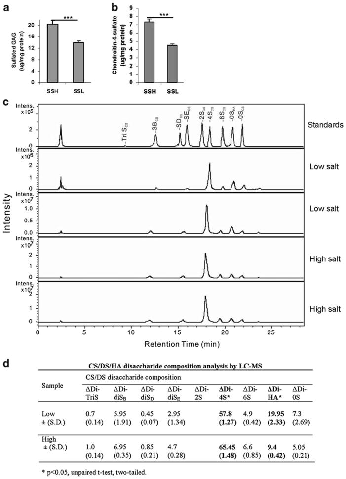
Increased sulfated GAGs, C4S, and sulfated disaccharides in renal tissue following higher salt exposure. a. sGAG in the rat kidney tissue following high salt was significantly greater in the SSH than in the SSL rats (p<0.0001, unpaired t-test, two-tailed; n=10 for SSH and for SSL). b. Similarly, the content of C4S in the rat kidney tissue was greater in the SSH rats with lower ARSB activity than in the SSL rat tissue (p< 0.0001; n=5 for SSH and n=5 for SSL). [ARSB = arylsulfatase B; GAGs = glycosaminoglycans; C4S = chondroitin-4-sulfate; SSH = salt-sensitive on high salt diet; SSL = salt-sensitive on low salt diet] c. 2-Aminoacridone (AMAC)-labeled chondroitin sulfate (CS)/dermatan sulfate (DS)/hyaluronan (HA) disaccharide analysis was performed by LC/MS. Chromatogram depicts the intensity of the disaccharides obtained following isolation, purification, and depolymerization of the glycosaminoglycans from the SSH and SSL renal tissue, and in comparison to standards. d. The disaccharide composition indicates marked differences between the average disaccharide content of the high and low salt renal tissue. The hyaluronan-derived disaccharide (ΔDi-HA) and the C4S-derived disaccharide (ΔDi-4S) are both present in high concentration and differ significantly between the high and low salt tissues (p=0.02 for ΔDi-HA and p=0.03 for ΔDi-4S, unpaired t-test, two-tailed). [CS = chondroitin sulfate; DS = dermatan sulfate; HA = hyaluronan; LC = liquid chromatography; MS = mass spectrometry; S = sulfate; UA = uronic acid; GalNac = N-acetylgalactosamine; GlcNAc = N-acetylglucos-amine; ΔDi-TriS = ΔUA2S-GlcNS6S; ΔDi-0S = ΔUA-GalNAc, ΔDi-4S = ΔUA-GalNAc4S, ΔDi-6S = ΔUA-GalNAc6S, ΔDi-UA2S = ΔUA2S-GalNAc, ΔDi-diSB = ΔUA2S-GalNAc4S;ΔDi-diSD =ΔUA2S-GalNAc6S; ΔDi-diSE = ΔUA-GalNAc4S6S; ΔDi = HA = ΔUA-GlcNAc; Intens = intensity]

