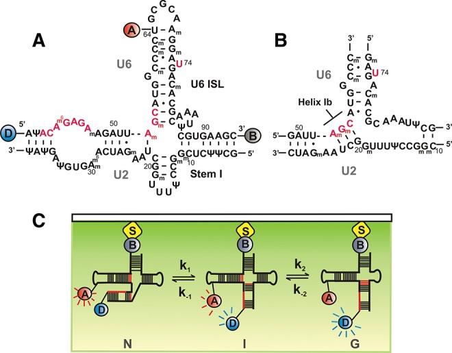
FIGURE 1.

hU2–U6 snRNA complex and single-molecule experimental setup. (A) Proposed four-helix structure of the hU2–U6 complex with FRET donor-D (Cy3, blue), FRET acceptor-A (Cy5, red), and biotin-B (gray). Highly conserved regions of U6 snRNA (ACAGAGA, AGC triad, and U74) are shown in red. (B) Proposed three-helix structure of the hU2–U6 complex containing helix Ib. (C) Total internal reflection fluoresence (TIRF)-based single-molecule experimental setup. Flurophore-labeled RNA was surface immobilized via a biotin-streptavidin linkage and excited using a 532-nm laser beam. Fluoresence intensities of donor (blue) and acceptor (red) fluorophores are collected through the objective and detected using a CCD camera.
