Figure 1.

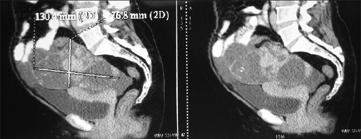
Computed tomography scan of the abdomen and pelvis revealed a 13 × 7.6 cm heterodense adnexal mass with solid and cystic components and punctate calcifications

Computed tomography scan of the abdomen and pelvis revealed a 13 × 7.6 cm heterodense adnexal mass with solid and cystic components and punctate calcifications