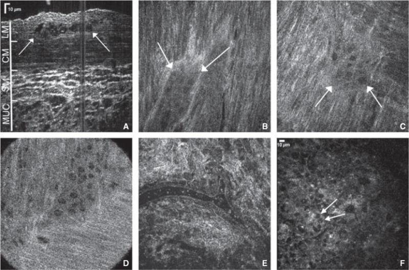
Figure 1.

Full-field optical coherence microscopy provides high resolution full-thickness imaging of gut wall. Cross-sectional reconstruction of stacked planar images obtained from the serosal side of the mouse colon reveals full-thickness colonic wall structures (A, arrows depict myenteric ganglion). Myenteric ganglia are visualized in gastric fundus (B), duodenum (C), and proximal colon (D). A colonic submucosal blood vessel containing erythrocytes is seen (E), as are crypts in the colonic mucosa (F, arrows mark goblet cells in the colon epithelium). LM, longitudinal muscle; CM, circular muscle; SM, submucosa; MUC, mucosa.
