
Figure 4.
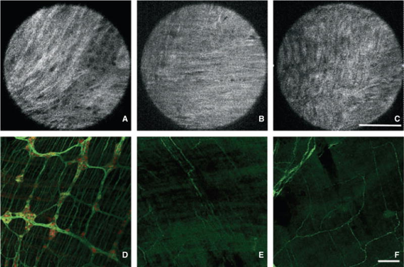
Morphologic assessment of enteric ganglia in Hirschsprung’s mice by Full-field optical coherence microscopy (FFOCM) and whole-mount immunohistochemistry (IHC). Representative images from FFOCM (imaged from the serosal side) and PGP9.5 (green)/Hu (red) whole-mount IHC were obtained from the proximal colon (A,D), mid colon (B,E), and rectum (C,F) of EdnrB−/− mice. The proximal colon is hypoganglionic while the distal segments have no visible ganglia in the images shown. FFOCM images are representative ‘real’ single images. Confocal microcopy images are shown with larger field of view in order to provide representative images of a large gut segment. Scale bars in C and F are 100 μm.