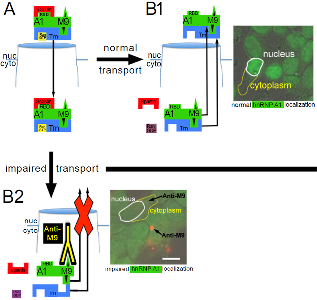
Figure 2. hnRNP A1 nucleocytoplasmic transport.
(A) Under normal conditions, hnRNP A1 (A1) binds transportin (Trn) via its M9 nucleocytoplasmic transport sequence; spastin RNA binds RNA binding domains (RBD) and upon RanGTP binding to Trn, the complex is transported through the nuclear pore to the cytoplasm (cyto). (B1) spastin and RanGTP are released into the cytoplasm, RanGTP is converted to RanGDP and hnRNP A1 and transportin return to the nucleus (nuc). The photomicrograph of SK-N-SH neurons shows that the vast majority of hnRNP A1 (green staining) is in the nucleus [118]. (B2) Under pathologic conditions when anti-M9 antibodies are present in neurons, hnRNP A1 transport from the cytoplasm to the nucleus is inhibited, thus hnRNP A1 is present equally in both the nucleus and cytoplasm. This may result in abnormal metabolism or transport of spastin and inhibition of binding of hnRNP A1 to Trn because of steric hindrance of the anti-M9 antibodies. The photomicrograph shows hnRNP A1 staining in the nucleus and cytoplasm, as well as fuorescently labeled anti-M9 antibodies (red) present in the neuronal cytoplasm [118].

