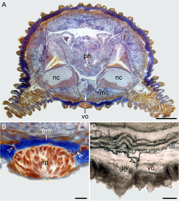
Figure 6.
Position and histology of the ventral and preventral organs in adult onychophorans. Light micrographs. Dorsal is up in all images. (A) Histological cross-section (Azan staining) at the level of a ventral organ in Metaperipatus blainvillei (Peripatopsidae). Note the thick layer of extracellular matrix (arrow) separating the ventral organ from the remaining tissues. (B) Detailed view of a ventral organ in Metaperipatus blainvillei. Arrowheads point to bundles of tracheal tubes that cross the extracellular matrix in the vicinity of the ventral organ. (C) Sagittal Vibratome section at the level of the ventral and preventral organs in Epiperipatus sp. 2 (Peripatidae). Anterior is left. Note the bundles of tracheal tubes (arrowhead) that open to the exterior next to the ventral and preventral organs. Abbreviations: em, extracellular matrix; ep, epidermis of the ventral organ; mc, median commissure; nc, nerve cords; ph, pharynx; pv, preventral organ; vo, ventral organ. Scale bars: 200 μm (A), 25 μm (B), and 50 μm (C).

