Figure 7.
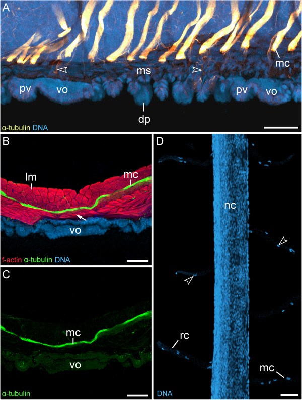
Median commissures and their spatial relationship to the ventral and preventral organs. Confocal laser-scanning micrographs of Euperipatoides rowelli (Peripatopsidae). (A) Sagittal section of a newborn juvenile, double-labelled with an anti-acetylated α-tubulin antibody (glow-mode) and a DNA marker (RedDotTM2; light-blue). Anterior is left and dorsal is up. Note that the serially repeated median commissures are not connected to the ventral and preventral organs but are spatially separated from them. Arrowheads point to single nerve fibres that innervate the dermal papillae. (B) Vibratome cross section of ventral body wall from an adult triple-labelled with an anti-acetylated α-tubulin antibody (green), a DNA marker (RedDotTM2; blue) and an f-actin marker (phalloidin-rhodamine; red). Dorsal is up. Note the layers of diagonal and ring musculature between the ventral organ and the median commissure (arrow). (C) Same section as in B showing only the anti-acetylated α-tubulin immunolabelling. Note the lack of nerve fibres or any connections between the ventral organ and the median commissure. (D) Detail of a dissected nerve cord from an adult specimen labelled with the DNA marker SYBR® Green showing “glial” cells that accompany the ring commissures and the median commissures (arrowheads). Lateral is left. Abbreviations: dp, dermal papilla; lm, longitudinal musculature; mc, median commissure; ms, musculature; nc, nerve cord; pv, preventral organ; rc, ring commissure; vo, ventral organ. Scale bars: 50 μm (A), 100 μm (B, C), and 50 μm (D).

