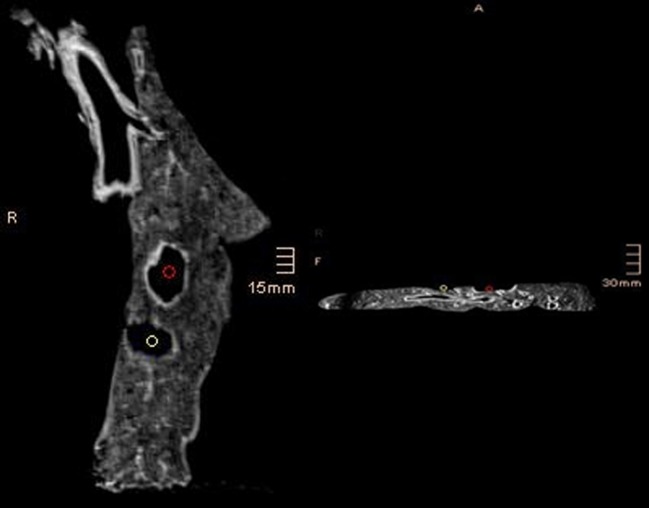
Figure 4:
Magnetic resonance tomography [T2 weighted, TE: 1.21, TR: 5.33, SL: 1, FA: 8] examination of the lung tissue after laser (red button) and monopolar cutter (white button) resection. After laser resection, the resection area revealed a 2–3 mm wide, hyper-intense, sharply delineated border (red button). No signal change was noticed in the adjacent lung tissue. The monopolar cutter created an irregular border of various depths (white button). The local tissue revealed areas that were sometimes more, other times less, hyper-intense at T2-weighting indicating a severe tissue damage.

