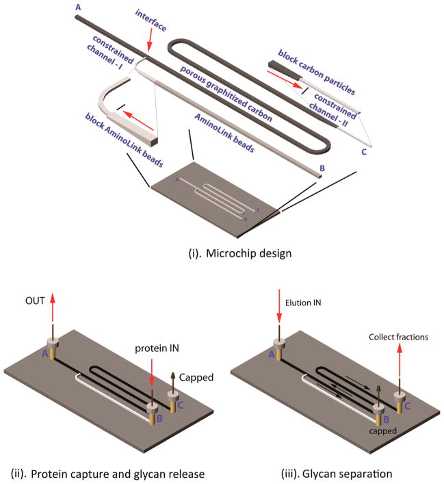
Figure 1.
GIG-chipLC for glycan analysis. The schematic diagram of the GIG-chipLC for glycan capture and separation and the location of A, B, and C for reservoirs and needle insertion. Two constrained channels (I and II) were constructed to block Aminolink beads and porous graphitized carbon particles (i); capture of proteins by infusing the proteins into the Aminolink bead-packed channel (ii); separation of released glycans in porous graphitized carbon particles (iii).

