Figure 2.
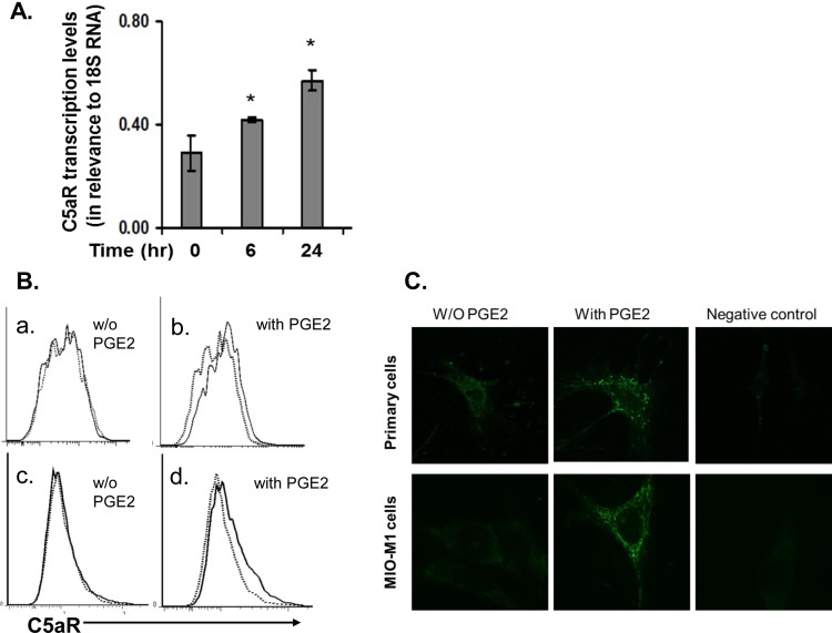
C5aR expression in Müller cells is upregulated by PGE2 as examined by qRT-PCR (A), flow cytometry (B), and confocal microscopy (C). (A) Quantitative RT-PCR assessment of C5aR expression in Müller cells after PGE2 stimulation. Müller cells were treated with 1 μM PGE2, and total RNAs were isolated after 0, 6, and 24 hours of PGE2 stimulation. C5aR transcript levels were measured by qRT-PCR and normalized against 18S RNA levels. (B) Flow analysis of C5aR expression on surface of Müller cells. C5aR was barely detectable in both primary cells (upper panel) and MIO-M1 cells (lower panel) under normal culture conditions (without PGE2); C5aR expression was increased and detectable in both types of cells after incubation with 1 μM PGE2 for 24 hours (with PGE2). Dotted line, isotype control; solid line, anti-C5aR mAb staining. Representative results from three individual experiments. (C) Confocal analysis of C5aR expression on Müller cells, showing results comparable to those from flow cytometry analysis.

