
Figure 2.
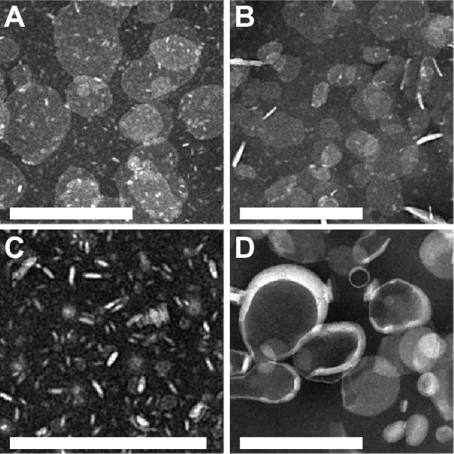
(A–D) Negative-stain transmission electron micrographs of NanoDisk–amphotericin B (ND-AMB) and liposomal AMB (L-AMB). (A) ND-AMB72C, (B) ND-AMB35C, (C) ND vehicle, and (D) L-AMB imaged by electron microscopy using magnification of 12,000× or 20,000×. Bar =200 nm.