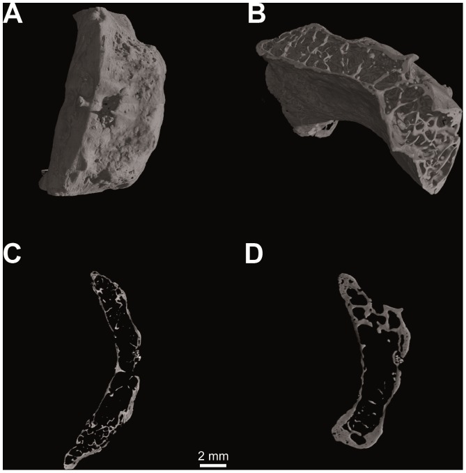
Figure 3. Computed tomography of Homo sapiens (N. S36-Sulmona Fonte d′Amore T64, University Museum Chieti - Italy).
Hyoid body volume rendering (V = 80 kV, I = 100 µA; pixel size: 12.5 µm; exposure time: 2.0 sec.; 2400 projections over 360 degrees) (a); spongy bone structure (b); histological architecture: medial sagittal section (c) and medial transverse section (d).

