
Fig. 4.
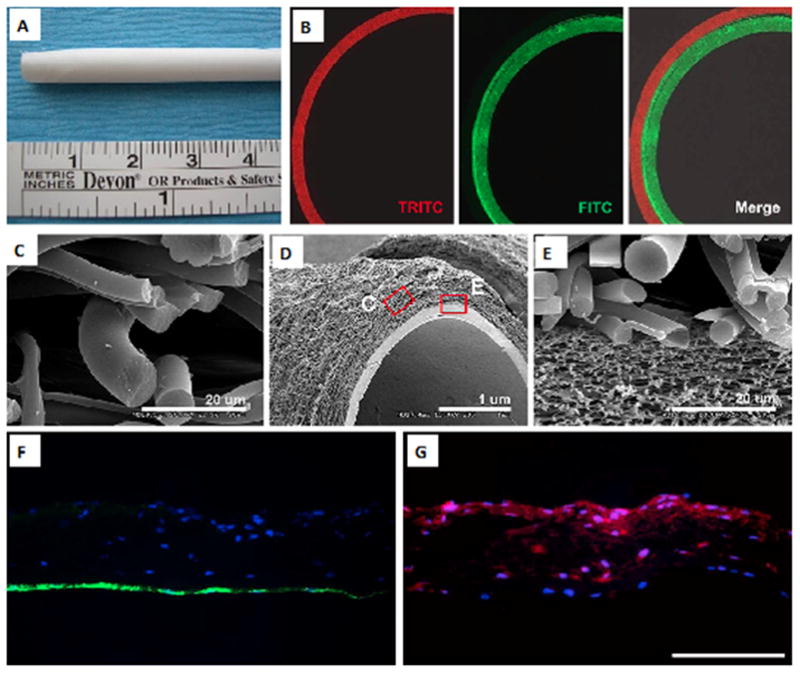
A PCL/collagen bilayer electrospun scaffold. (A) Macrostructure of the scaffold; (B) fluorescent images of the scaffold; (C–E) SEM images of different layers of the scaffold, (C) outer layer, (D) bilayer structure, and (E) interface between the inner and outer layers; (F–G) fluorescent images of EC and SMC seeded scaffold (F) EC seeded inner layer (green: CD31 expression), indicating the formation of an EC monolayer and (G) SMC seeded outer layer (red: α-SMA expression), demonstrating SMC infiltration into the outer layer (scale bar in F and G: 500 μm). Reproduced from [57] with permission from Elsevier.