
Fig. 5.
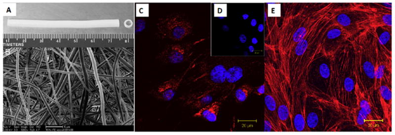
An electrospun tubular scaffold fabricated using recombinant human tropoelastin (rTE), (A) Front view showing the length of the scaffold (7 cm in length, 4 mm inner diameter), (B) The random orientation of the rTE fibers, average diameters: 580 ± 94 nm (scale bar: 5 μm); (C) vWF (red) staining and DAPI (blue) nucleus staining, (D) vWF and DAPI staining of the control sample, (E) The EC monolayer fromed on rTE scaffold (15 wt.%) imaged after 48 hours of culturing. The cytoskeletal actin fiber was stained with rhodamine phalloidin (red) and the nuclei were stained using DAPI. Reprinted from [10] with permission from Elsevier.