
Fig. 7.
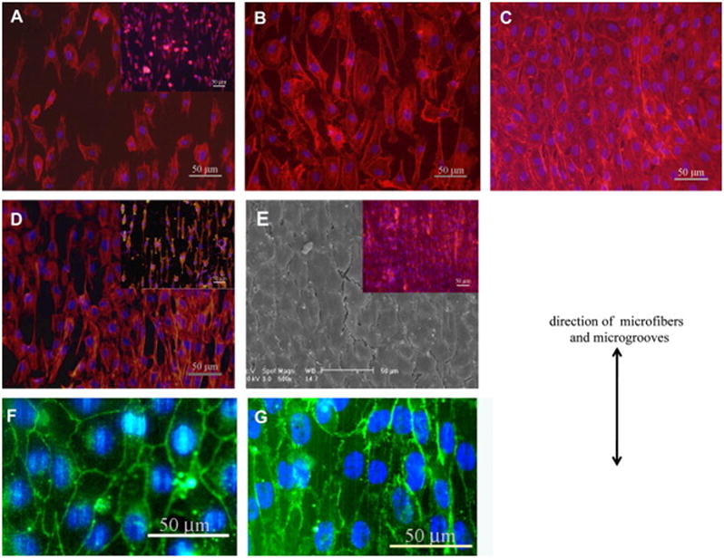
Endothelial cell morphology on the micropatterned scaffold: (A–C) adhesion and spreading of cells inside microfiber patterned electrospun graft. Fluorescently stained cell nuclei (blue) and actin filaments (red) of BAECs or EA.hy926 cells (inset) at (A) 2 hours, (B) 3rd day, and (C) 5th day after seeding. (D, E) adhesion and spreading of cells inside microgrooves patterned hybrid grafts. (D) Fluorescence image of BAECs or EA.hy926 EC (inset) on 3rd day after seeding. (E) The confluent monolayer of BAEC and overlaid fluorescence image of a EA.hy926 EC monolayer (inset). (F, G) VE-cadherin staining (green) of (F) BAEC and (G) EA.hy926 monolayers on microgrooves on 7th day after seeding. The direction of microfibers and microgrooves is shown using the double-headed arrow. Reprinted from [53] with permission from Elsevier.